

Applying AI in Radiology to Optimize Workflow for Chest X-rays
Reading Time: 8 minutes read
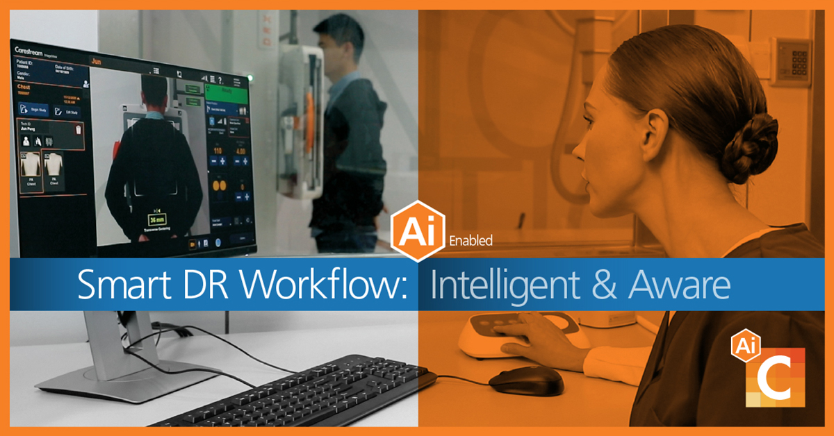
Smart DR Workflow helps improve image quality and consistency, and speed patient throughput.
Applications for Artificial Intelligence (AI) in radiology are increasing, especially in workflow where they can help streamline exam efficiency and provide more consistent outcomes. Carestream’s software powered by Eclipse uses AI to optimize exam workflow while improving diagnostic image quality and consistency for the acquisition of a Chest PA view. Our AI-based Smart DR Workflow has 3 important components:
- Smart Positioning
- Smart Technique
- Smart Collimation
In this blog, I explain how we are currently leveraging AI in radiology for the acquisition of a Chest PA view.
AI for DR Workflow for Chest PA view
Knowledgeable radiographers understand that correct positioning and accurate settings of X-ray equipment are essential to capturing an image that will meet diagnostic requirements. However, executing these steps require precision and time, and even the most skilled radiographers can sometimes fail to obtain clinically viable results. (1)
For example, prior to making the exposure in a Chest PA view, the radiographer must guide the patient into the correct position, adjust the collimation and set the proper exposure technique according to the patient’s size. Another example: the radiographer might use improper positioning or incorrect exposure parameter settings, resulting in an image that fails to meet diagnostic requirements.
Our new AI-based Smart DR Workflow helps the radiographer acquire images in a consistent manner from patient to patient, enabling more consistent presentation and image quality, and reducing retakes. It makes DR systems more “intelligent and aware” of the environment and patient by integrating sensors, cameras and AI software. It simplifies the workflow of a Chest PA view acquisition by offering audio and video guidance, as well as assistance in adjusting Bucky height, positioning the patient, and setting technique and collimation size. Let’s look deeper at how Carestream is applying AI in radiology through the three components of Smart DR Workflow.
Smart DR Workflow: Smart Positioning
Smart Positioning utilizes AI to evaluate the overall patient body position to ensure both equipment and patient positioning meet clinical examination requirements. Two RGBD (RGB & Depth) cameras collect information that is sent to a pose-detection algorithm and a classifier. These additional hardware components also are required: two controllers, a hub, a console PC, markers for recognition, and indicators of examination and preparation areas.
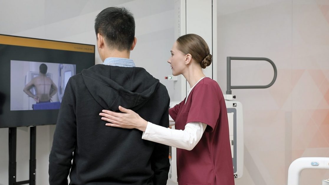
A Video Assist display is located in the examination room to provide the patient with exam information along with a picture illustrating how to position themselves next to the equipment. Once the patient moves to the preparation area, the required Bucky height is automatically calculated along with verification that no collision caused by Bucky movement can occur. When the Bucky has moved to the proper height, the patient can position themselves next to the Bucky as indicated on the Video Assist display. Correct positioning information will be checked with any error and required adjustment presented to the radiographer. This includes shoulder-height, patient contact with Bucky, pose, center alignment, orientation, tilt, and hand position.
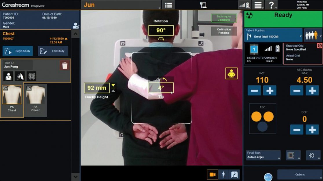
All of these indicators can be seen on the console display. All the while, the radiographer is in full control and can override an operation and/or take an exposure at any time, even if patient positioning is not correct.
I’d also like to mention Audio Assist, even though it is not based on AI technology. This separate feature can be customized with pre-recorded audio clips according to the facility’s needs. It can also allow direct, real-time voice communication to the patient. Audio Assist also provides patient safety recordings, for example, alerting a patient prior to automatic movement of the Bucky.
The Smart Positioning capability allows the radiographer to correct positioning errors prior to acquisition, thereby reducing the time, cost and additional dose associated with a repeat exposure while improving image consistency. Additionally, Smart Positioning can provide a valuable training tool for less experienced radiographers. Its automated features also should decrease the time that a radiographer needs to be in close contact with patients for positioning — a huge benefit in the presence of infectious diseases.
Smart Technique
Exposure technique (ET) is the key factor to achieving image quality in a DR imaging system. In the current DR market, auto exposure control (AEC) is widely used to control the exposure time at a given dose. Nonetheless, due to hardware limitations, AEC cannot be used in mobile or fixed tabletop DR — leaving default ET or manual adjustment as the only options. Because default ET is not suitable for every patient, radiographers must take the extra time to adjust the ET — which depends on many variables including diagnostic factors, patient factors, image chain factors and special factors.
Setting ET too high or low leads to overexposure and underexposure, respectively. The former exposes the patient to unnecessarily high levels of radiation, while the latter results in poor image quality [2,3].

To help address this, our AI-driven Smart Technique automatically detects the patient’s size to apply proper ET. This minimizes the need for the radiographer to do manual adjustments thereby reducing their workload and helping to lower patient radiation dose and ensure image quality. It uses an RGBD camera to capture patient information, and applies AI algorithms to detect patient thickness, shoulder width and upper body height to calculate patient size. Then it automatically determines ET according to patient size, the area of interest and diagnostic requirements.
Smart Collimation
The current collimation workflow in DR radiography requires the radiographer to adjust the collimator’s settings manually to match the different body parts to be imaged. This is problematic for two reasons. First, it requires time. Second, the collimator adjustments made by individual radiographers based on their differing levels of expertise and subjective visual measurements will be disparate. This may lead to a higher-than-necessary dose of radiation for some patients — especially those imaged by less-experienced radiographers [4,5].
By applying AI to radiology, we developed Smart Collimation capabilities. Smart Collimation can automatically adjust the collimator blades to the appropriate field size for different patients, reducing both the radiation dose to patients and the workload of radiographers — freeing them to provide a higher level of care. In addition, the appropriate collimation field size can reduce the influence of X-ray scatter and improve image quality.
Smart Collimation utilizes camera data to recognize the human shoulder joint, then calculates shoulder width and height to determine the correct collimation field size and settings. The system can determine correct collimation height (the length of collimation in the vertical direction) for Chest PA based on patient shoulder height and detector size.
After the patient is correctly positioned, the system also can determine correct collimation field width (the length of collimation in the horizontal direction) based on the patient shoulder width and the detector size.
The significant advantages of Smart Collimation are the potential reduction of patient dose and improvement in radiographer productivity.
Benefits of AI in Radiology Workflow
Medical imaging systems can fail to generate qualified diagnostic images for many reasons, including incorrect positioning of the patient and/or the system. Applying AI to automate many of the steps in image acquisition helps to achieve correct positioning and accurate settings of X-ray equipment. These AI workflow features help increase exam throughput, adding revenue to the bottom line; and give your radiographers added confidence and job satisfaction. Equally important, being able to perform an exam in less time and with less movement by the radiographer can increase satisfaction for both the patient and the radiographer. #carestreamAI #radiologyAI
About the author: Lei (Leo) Sun(Ph.D.) is a senior image quality engineer at Carestream Health. He focuses on the optimization of image quality, workflow and dose in digital radiography.
Learn more about how Carestream is applying AI in radiology:
- Smart Noise Cancellation
- AI Features in Radiology to Adopt Today
- Eclipse Intelligence in Action
- The “Why” Behind Applying AI in Radiology
References:
- Sajjad Rastegar, Jalal Beigi, Ehsan Saeidi, Reject analysis in digital radiography: A local study on radiographers and students’ attitude in Iran, Med J Islam Repub Iranv.33; 2019PMC6708103
- Exposure Issues, https://www.upstate.edu/radiology/education/rsna/radiography/issues.php
- J. Anthony Seibert, Richard L. Morin, The standardized exposure index for digital radiography: an opportunity for optimization of radiation dose to the pediatric population, Pediatr Radiol. 2011; 41(5): 573–581, doi: 10.1007/s00247-010-1954-6
- Ofori K, Gordon SW, Akrobortu E, Ampene AA, et al Estimation of adult patient doses for selected X-ray diagnostic examinations. J Radiat Res Appl Sci. 2014; 7(4):459-62.
- Engel-Hills P. Radiation protection in medical imaging. Radiography. 2006;12(2):153-60.
- Tested Smart Positioning performance in Renji hospital (Shanghai, May 2020) and The First People’s Hospital of Kunshan (May 2020). Smart Positioning demo placed in these two hospitals for clinical trials for one week.


Lovart
Wow, this Smart DR Workflow sounds super helpful! Less retakes and better image quality? That’s a win-win for both patients and radiographers. AI in radiology is definitely the future! By the way, if anyone’s interested in exploring AI-powered design tools, I’ve been using an AI design agent called Lovart (https://lovart.ai) and found it really helpful for creative projects.