

Improving the Quality of Mobile Chest X-rays
Reading Time: 7 minutes read
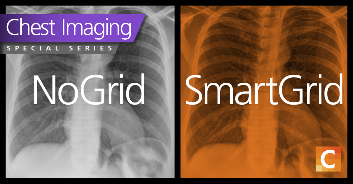
Compensating for the effects of X-ray scatter improves contrast.
Mobile chest X-rays are often performed on acutely ill patients who are too critical or unstable to be transported to an X-ray imaging room. For these patients, bedside mobile X-rays can be the only option for medical imaging.
Fortunately, in recent years, advances in mobile imaging have come a long way. Many imaging professionals tout today’s portable systems as “X-ray rooms on wheels” that help eliminate the risks, costs, and delays of patient transport by bringing diagnostic technology directly to the bedside.
Recognizing the disadvantages of mobile imaging
Yet, mobile imaging still has potential drawbacks. In speaking about X-ray studies performed with mobile systems, Dr. David Levin, professor and chairman emeritus of the Department of Radiology at Thomas Jefferson University Hospital in Philadelphia, had this to say. “If you compare the quality of those studies with the quality of a study that was performed in a hospital in a radiology department or in a private radiology office, there is going to be no comparison,” he told Reuters Health. “If a portable X-ray is absolutely necessary because of the patient’s clinical condition, then it’s justifiable.”(1)
Other variables adversely affecting portable imaging, according to physicians Leif Jensen and Christopher Meyers, include “… patient factors such as obesity, hypoventilation, and motion unsharpness.” (2)
Difficulties of mobile chest X-rays
Concern over mobile image quality is perhaps greatest when it comes to X-rays of the chest. Ideally, chest studies are performed with the patient in standing position. This is important for several reasons. One, it prevents blood engorgement of pulmonary vessels, which can compromise image clarity.
Two, upright positioning permits better visualization of air and fluids in the chest. When the patient is imaged while supine, fluids can diffuse themselves across the surfaces of the lung, producing a hazy image. So mobile imaging can be inherently problematic. If a patient must be X-rayed while lying in bed, the above-mentioned factors can compromise image quality from the get-go.

The issue of radiation scatter in mobile chest X-rays
Further complicating the situation is the potential degradation of image quality due to radiation scatter. This phenomenon occurs most frequently when imaging thicker areas of the body – such as the chest – particularly if collimation is not in close enough proximity. Specifically, when the X-rays penetrate the chest, a percentage of the photons engage in Compton interactions and cause radiation to scatter. This compromises image quality by introducing a noise-laden, low frequency background signal that creates a haze. The result is an image with reduced contrast and detail, creating the potential for obscured vasculature, infiltrates and other pathology. (3)
Are grids the best answer to improve chest X-ray quality?
The traditional solution to reduce scatter has been the use of anti-scatter grids. Grid design includes parallel strips made of lead as well as strips composed of a radiolucent material. The technologist situates the grid in between the detector and patient. The beam, its path parallel to the radiolucent strips, passes freely between them. Radiation scatter is largely blocked by the angled lead strips before it can reach the detector and compromise the image. This helps preserve clarity and diagnostic value.
Grids can be highly effective in reducing scatter. But, it seems every solution has its disadvantages, and that is true here as well. Grids typically require a higher dose of radiation exposure, as the X-ray beam is attenuated by the lead strips. Also, grids are heavy and bulky. This can lead to misalignment during positioning, which can reduce the grid’s efficacy. And, because they are inconvenient to handle, radiologic technologists might be discouraged from using grids altogether in some situations.
SmartGrid provides image quality comparable to images acquired with an anti-scatter grid
Recently, Carestream introduced software that reduces the damaging effects of scatter radiation in an image – helping to improve the contrast of the image when a physical anti-scatter grid is not used. Carestream’s software, called SmartGrid, uses an advanced algorithm that estimates low-frequency scatter distributed throughout an image and reduces it.

Figure B (center): Same patient, same SID @ 95 kVp, 2.8 mAs, no Grid, processed with SmartGrid;
Figure C (right): Same capture as B, without SmartGrid. Click through to see a higher-resolution image.
Many physical factors affect the properties of scatter including energy spectrum of the beam, thickness and size of the object, and collimation. But by using empirical modeling, SmartGrid software can accommodate these factors through estimation of algorithm parameters tuned to replicate anti-scatter grid visual performance.
SmartGrid is available for use with Carestream’s flagship DRX-Revolution Mobile X-ray System and scaled-down affordable DRX-Revolution Nano. (Available for commercial sale in the US and Canada.) Dates in European Union will be announced shortly.) SmartGrid also can be used with mobile systems from other manufacturers upgraded to digital with Carestream’s DRX Retrofit Kits. In addition, it is available in all our modalities. This makes it a highly effective solution to the issue of compromised image quality in mobile chest X-exams.
SmartGrid processing provides image quality comparable to images acquired with an anti-scatter grid lowering radiation dose in bedside chest imaging. The benefits of grid-like image quality without the use of an anti-scatter grid can lead to improved workflow and ease of imaging for radiographers, producing a win-win for a busy hospital.

Learn more:
- Read the white paper below or download it here to learn more about SmartGrid technology and the clinical study evaluating paired grid and non-grid portable chest images at the University of Rochester Medical Center.
- Read Liverpool Heart & Chest Hospital’s Guide to Mobile Chest X-rays for Thoracic and Cardiac Care.
- Read about the University of Utah COVID Chest Imaging Through Glass.
References
- Reuters Health News:Portable X-ray services becoming more common https://www.reuters.com/article/us-portable-xrays/portable-x-ray-services-becoming-more-common-idUSKBN0KW1ZN20150123
- Applied Radiology: Reducing Errors in Portable Chest Radiography https://appliedradiology.com/articles/reducing-errors-in-portable-chest-radiography
- Perry Sprawls, Ph.D., Scattered Radiation and Contrast http://www.sprawls.org/ppmi2/SCATRAD/


Lori Barski and Mary Couwenhoven are imaging scientists in the Carestream Health Research and Innovation Image Processing & Analysis group. Dong Yang also was a member of the Image Processing and Analysis Group during the research phase of the SmartGrid project.


Luis Viana
I like to know how much does cost a X-ray grid for a portable X-ray ray CR machine
Kathleen Remis
Hello, thank you for your interest. I sent an email with information to your gmail address. You can also find a local contact in your area by visiting this page https://www.carestream.com/en/us/services-and-support/world-wide-contacts#?cat=Medical
Kathleen Remis
Hello Luis, kindly go to this page where you can find a helpful contact in your area https://www.carestream.com/en/us/services-and-support/world-wide-contacts#?cat=Medical