

White Paper: Addressing Pediatric Imaging Challenges with Carestream Solutions
Reading Time: 11 minutes read
Learn the techniques behind maximizing dose efficiency for pediatric imaging.
Table of contents
- Pediatric Solutions for Image Acquistion
- Pediatric Solutions for Image Processing
- Pediatric Solutions for Image Review and Assessment
Pediatric Solutions for Image Review and Assessment
Best practices of radiographic imaging follow the principle of using a dose “as low as reasonably achievable” or ALARA,(1) which balances the needs of the patient (lower dose) with the necessity of producing an image with quality suitable for confident exam interpretation. While dose level is a major aspect of managing dose efficiency for any patient, radiographic imaging of pediatric patients presents several unique challenges. Increased radiation sensitivity of growing organs and bones, children’s longer expected lifespans and the large range of body habitus encompassed by this patient demographic all mean that it’s not appropriate to use the same acquisition techniques and image-processing parameters used for adult imaging. The Image Gently campaign’s “Back to Basics” initiative encourages the use of pediatric-specific imaging practices and is completely consistent with the guiding principles in Carestream’s approach to these important issues. (2, 3, 4)
To provide the highest quality image with the most efficient use of the radiation exposure, it’s important to address each step in the image-formation chain as part of a complete system. The image-formation process can be naturally divided into three distinct stages: image acquisition, image processing for display, and image review and assessment. In this blog, I’ll review at a high level how Carestream is addressing pediatric imaging challenges throughout the image formation process: image acquisition, image processing for display, and image review and assessment. For a deeper dive, read our white paper.
Pediatric Solutions for Image Acquisition
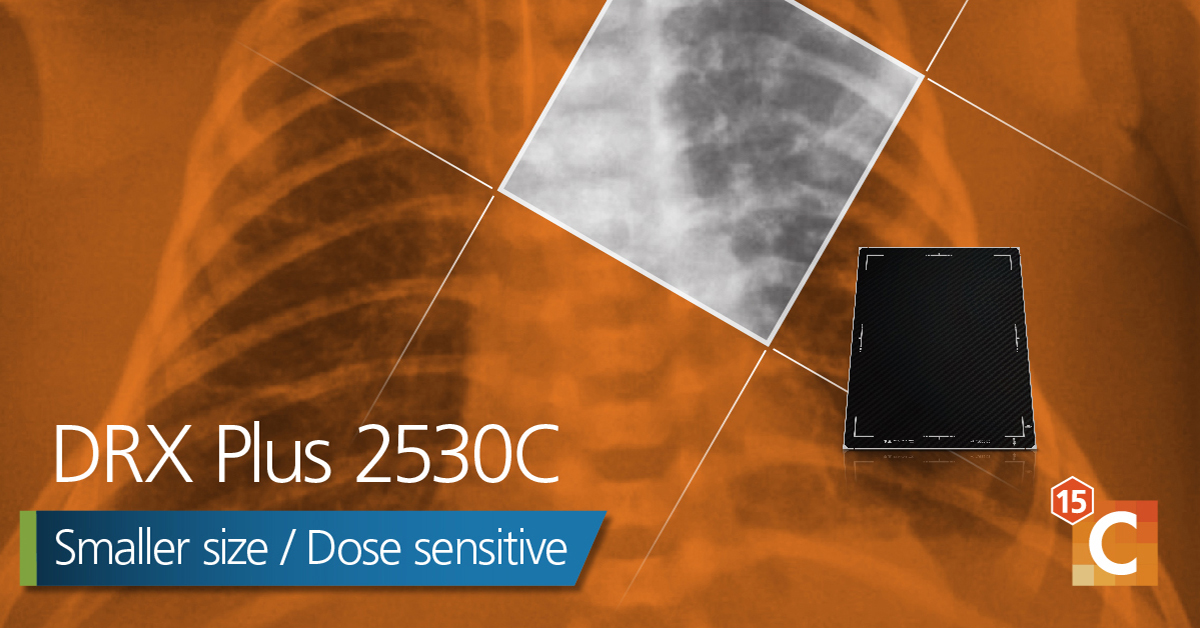
Image Receptor: Capturing the X-ray image with the image receptor is the first stage of image formation. The introduction of Carestream’s wireless DRX detector products has been a major step forward in the provision of a high-quality X-ray detector that fits seamlessly into the workflow of the NICU and pediatric ICU. In addition, the use of a cesium scintillator layer helps to ensure the best possible image quality. The design virtually eliminates the problems that can be encountered with patient positioning in a busy clinical environment when a tethered system is used. The replaceable battery also guarantees that the detector is ready for use at a moment’s notice. The CARESTREAM DRX Plus 2530C panel is a small-format, high-resolution (98 µm pixel spacing), high-detective quantum-efficiency (DQE) panel that fits easily into a neonatal incubator X-ray tray and is ideal for tabletop extremity exams.
Appropriate Acquisition Techniques: In addition to using a highly efficient detector, it’s also essential to use the appropriate acquisition techniques (e.g. kVp, mAs and filtration) across the wide range of pediatric body habitus. This range of body sizes – from the smallest neonatal patient to the largest adolescent – requires acquisition techniques to be tailored to each patient’s size and age. To help with this challenge, Carestream offers the ability to select the pediatric patient body size (optionally based on weight or age) from a range of seven categories, which is an expansion of size categories recommended by the FDA. (5, 6)
Pediatric Capture Optimization and Enhancement: This option allows the system to choose default acquisition parameters and image-processing configurations appropriate for different sizes of patients and different detector types. This capability provides a more consistent acquisition and display of images for patients within a given body size and age range.
Smart Noise Cancellation: One of the most significant recent Carestream advancements is Smart Noise Cancellation (SNC), which has a direct bearing on the selection of acquisition techniques. This new AI-based denoising technique facilitates dose reduction across the board for all patient sizes and general radiography exams while preserving fine spatial detail. (7)
Carefully designed reader studies have demonstrated that with a cesium iodide panel, acquisitions at 800 ISO speed with SNC applied were rated superior in image quality when compared to 400 ISO speed acquisitions without the use of SNC. (7) Likewise, for gadolinium oxysulphide (GOS) panels, acquisitions at 500 ISO speed were deemed superior when compared to corresponding 320 ISO speed exams without SNC. Overall, 99% of the low-dose image pairs with SNC were rated as good or better than the nominal dose images without SNC.7 (Note: ISO speed has an inverse relationship to IEC exposure index. As exposure is halved, IEC EI is halved, whereas ISO speed is doubled.) And when SNC is coupled with other dose-management methods such as filtration, (8) even greater dose reduction can be achieved.
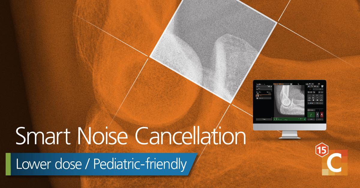
SNC significantly benefits image quality across all exams for both the DRX1 and DRX Plus family of detectors. SNC coupled with the best practices promoted in the Image Gently campaign can provide maximum image quality at significantly reduced dose.
DRX-LC Detector: Carestream now offers the DRX-LC detector that is specifically designed for long-bone and spine imaging using single-shot exposures that enable fast image acquisition and preview, simplified workflow and reduced dose compared to multi-shot long-length imaging. While it’s difficult to directly compare the multi-shot to single-shot LLI relative to radiation exposure, all things being equal (grid, SID, kVp and detector imaging performance), the multi-shot LLI method results in approximately 10% more dose overall and 100% more dose in the overlap anatomical regions. Considering the long hold time associated with multi-shot LLI, where patient motion frequently results in a repeat of the exam, the dose reduction from reduced repeats is also an important consideration.
SmartGrid Software: X-ray scatter can significantly degrade image quality if it’s not managed as part of the acquisition process. The use of an anti-scatter grid decreases the amount of scatter that reaches the imaging plate and improves image quality. But a major drawback of using a grid is the required increase in dose to the patient. Measuring patient thickness is highly recommended as part of selecting the optimal technique. Grids are appropriate for body part thicknesses greater than 12 cm, but in pediatrics, exceptions may be made for exams that contain a substantial amount of air, such as chest exams. (9)
Read the Complete White Paper on Addressing Pediatric Challenges
However, recent advances in image processing have replaced the need for using a physical grid with software-based scatter suppression. Carestream’s SmartGrid processing now makes it possible to image pediatric patients without a grid, thereby lowering the radiation exposure to these patients.
Prior Image Review: In the spirit of driving continuous improvement and consistency, Carestream’s Prior Image Review feature enables technologists to review prior acquisitions of the patient on the console screen. Priors are pulled back from PACS (even from other vendors’ equipment) as the radiographer is setting up the current exam. Previous positioning and technique factors can also be reviewed, enabling the technologist to learn and replicate the finer points of good previous studies. With the press of a button, the same techniques can be copied from a prior exam to the current exam, thus driving consistency between technologists.
Once an image has been acquired, rapid display of the preview image allows the radiographer to quickly decide whether the patient’s anatomy was correctly captured or if the image needs to be retaken. This improves the speed and efficiency involved in completing exams, which is particularly important for young patients. To help, Carestream provides the IEC Exposure Index (EI) for quick assessment of the amount of radiation used to create the image.(10, 11)
The associated Deviation Index (DI) allows an immediate evaluation of the acquisition technique compared to the institutional exposure target for the specific exam. This instant feedback, coupled with the other developments in technique selection described above, helps the radiographer provide more consistent image quality from the detector to the next step in the imaging chain – image processing.
Pediatric Solutions for Image Processing
EVP Plus image Processing and Display: Once a high-quality image has been acquired at the lowest possible patient exposure, it’s essential to perform appropriate image processing that presents the diagnostic information clearly and most efficiently to the radiologist. Carestream’s EVP Plus Software, powered by our Eclipse image-processing engine,(12) can be tailored to adjust the image-processing parameters to an individual site’s preference.
Pediatric Capture Optimization and Enhancement: When coupled with the Pediatric Capture Optimization and Enhancement option, the image processing parameters can also be adapted to display the clinical features in a more informative way compared to using adult image-processing configurations. The multi-band frequency decomposition, with either traditional noise reduction or SNC if enabled, and controlled edge-restoration capabilities mean that the available clinical content of the bony structures in the smallest NICU patients can be appreciated as well as the trabecular detail of older, more developed patients, as one example. The fine detail and lower contrast of the smallest NICU patient’s anatomy require accentuation of different frequency components than those of larger adolescents.
Tube and Line Visualization: Available for all patient sizes, Tube and Line Visualization uses a companion image automatically created from the original acquisition with optimized processing for clearer, easier visualization of lines. Examples include a peripherally inserted central catheter (PICC), central venous catheter, and tubes, such as nasal gastric (NG) and endotracheal (ET), which help to avoid repeat imaging.
Pediatric Solutions for Image Review and Assessment
Quality Acceptance and Control: Once an imaging system has been installed and tailored to a site’s preferences for patient exposure and image “look,” it’s important to have an ongoing quality control (QC) program in place that ensures the continued high quality of the images delivered to the reading radiologist. There are multiple aspects to this type of QC program and Carestream Health has implemented several system capabilities that enable a site to easily track many of the important parameters.
At the front end, the DR Total Quality Tool (DR TQT) package allows for efficient evaluation of the digital X-ray detector’s current performance level. In addition, the IEC EI allows quick evaluation of the exposure levels used to acquire the images.
On a departmental level, our Administrative Reporting and Analysis Software allows the QC technician or physicist to query all the Carestream systems across the institutional network from a single, central location. This can quickly highlight anomalous exposure levels, high repeat rates or other image-quality issues that may develop and provides opportunities to identify and resolve problems at an early stage. Together, these system capabilities can help technologists maintain their high level of image quality and consistency to facilitate reliable diagnoses. Carestream supports DICOM Radiation Dose Structured Reports, enabling institutions to track and monitor all irradiation events.
The unique demands of pediatric imaging require a systemwide approach to guarantee high-quality imaging at the lowest possible exposure for young patients. Carestream Health offers a range of features and functionality that ensure our systems can provide the best and safest possible X-ray imaging across the full range of clinical exams for all pediatric patients.

Lori Barski is an imaging scientist in the Data Science Group at Carestream Health. She is responsible for image processing development and image quality optimization. She holds 19 patents, and has worked with Kodak/Carestream Health for 40 years.
References:
1 Miller, Donald & Schauer, David. (2015). The ALARA principle in medical imaging. AAPM Newsletter. 40. 38-40.
2 Bulas DI, et al. AJR Am J Roentgenol. May 2009; 192(5):1176-8. Image Gently: Why We Should Talk to Parents about CT in Children.
3 AJR Am J Roentgenol. May 2009; 192(5):1169-75. Image Gently Vendor Summit: Working Together for Better Estimates of Pediatric Radiation
Dose from CT. Strauss KJ, et al.
4 Image Gently®: The Alliance for Radiation Safety in Pediatric Imaging. http://www.pedrad.org/associations/5364/ig/ (Accessed September 27,
2012).
5 FDA guidance entitled “Premarket Assessment of Pediatric Medical Devices,” May 14, 2004.
http://www.fda.gov/downloads/MedicalDevices/DeviceRegulationandGuidance/GuidanceDocuments/UCM089742.pdf.
6 FDA guidance entitled “Guidance for Industry and Food and Drug Administration Staff: Pediatric Information for X-ray Imaging Device
Premarket Notifications,” November 28, 2017. https://www.fda.gov/regulatory-information/search-fda-guidance-documents/pediatricinformation-
x-ray-imaging-device-premarket-notifications (Accessed July 12, 2022).
7 Smart Noise Cancellation Processing: Providing a New Level of Clarity in Digital Radiography and a Foundation to Reduce Dose.
https://www.carestream.com/blog/wpcontent/ uploads/2022/05/white_paper_smart_noise_cancellation_low_dose_2000299_202203_ltr_en_2.pdf (Accessed Jun 15, 2022).
8 Tugwell-Allsup JR, Morris RW, Thomas K, Hibbs R, England A. “Neonatal digital chest radiography – should we be using additional copper
filtration?,” Br J Radiol. February 1, 2022; 95(1130):20211026. doi: 10.1259/bjr.20211026. Epub December 14, 2021. PMID: 34797726; PMCID:
PMC8822573.
9 American Society of Radiologic Technologists. Best Practices in Digital Radiography. (2019). https://www.asrt.org/docs/defaultsource/
research/whitepapers/asrt12_bstpracdigradwhp_final.pdf?sfvrsn=743d0370_16 (Accessed June 28, 2022).
10 International Standard IEC 62494-1 (2008) Medical electrical equipment – exposure index of digital X-ray imaging systems – Part 1: definitions
and requirements for general radiography. International Electrotechnical Commission, ISBN 2-8318-9944-3.
11 Seibert J.A., Morin R.L., “The standardized exposure index for digital radiography; an opportunity for optimization of radiation dose to the
pediatric population,” Pediatric. Radiol. 41(5), (2011), 573-581.
12 https://youtu.be/4AS05QuGWd8 (Accessed July 13, 2022).


