Carestream Sign In
Welcome to Carestream.com's communities.
Our customers and partners have access to powerful online communities and tools. Use this overview to discover the best destination for you. Registration and sign-in are required to access these websites.
Vue Cloud Community
Carestream's Vue Cloud Community is your single point of access to the tools you need to diagnose exams, review a patient portfolio or view real-time department performance.

Carestream.com Communities and Tools
Welcome to Carestream.com's communities.
Our customers and partners have access to powerful online communities and tools. Use this overview to discover the best destination for you. Registration and sign-in are required to access these websites.
Vue Cloud Community
Carestream's Vue Cloud Community is your single point of access to the tools you need to diagnose exams, review a patient portfolio or view real-time department performance.

Taking Better Electron Micrographs
 Transmission electron microscopes (TEMs) provide images with extremely high resolution for specimen analysis. The challenge is to capture these images on film without sacrificing this level of detail. The very elements that make this detail possible-the electrons- contribute to the difficulties inherent in electron micrography.
By using electrons effectively, however, it is possible to optimize image quality by maximizing density, enhancing contrast, and reducing noise.
The key is to use more electrons: in other words, to increase the sampling rate.
Increase Exposure Time Reduce Magnification
Adjust Exposure and Development Causes of Electron Micrography Noise
Increase Exposure Time
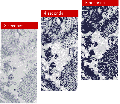
The simplest way to use more electrons to enhance image quality is to increase exposure time, as illustrated in Figure 2. Here, the obvious benefits are increased density and contrast. Additionally, the signal-to-noise ratio also improves: the image signal increases linearly with exposure (number of electrons absorbed), while the noise structure increases less rapidly with the square root of exposure. Of course, it is also possible to increase density and contrast by increasing development activity (e.g. by increasing development time), but signal and noise increase proportionately, leading to a less favorable signal-to-noise ratio than is achieved by increasing exposure time.

Figure 2: Specimen and instrument conditions permitting, increasing exposure will increase negative density, enhance image contrast, and improve signal-to-noise ratio.
Reduce Magnifcation
Some specimens cannot tolerate long exposure times due to their instability or other considerations. In these cases, a reduction in instrument magnification will produce better image quality. If needed, magnification can then be regained photographically, either with a loupe or through photographic enlargement.

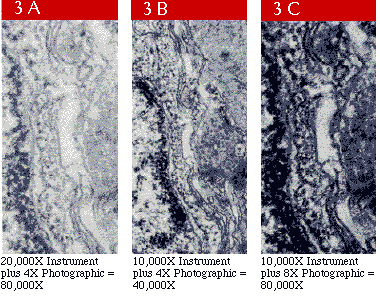
Figure 3: Reducing instrument magnification and compensating with little loss in fine detail (3C versus 3A). Results are comparable to a micrograph recorded at a lower total magnification (3B).
Reducing magnification while allowing all other conditions to remain the same permits more electrons to strike a unit area of emulsion on the film without changing the number of electrons passing through the specimen. As seen in Figure 3, the resulting micrograph is more dense and has a higher contrast (3B versus 3A). The magnification that is sacrificed can be regained through a correspondingly greater optical magnification (as illustrated in 3C). However, this restoration in image size may be accompanied by an increase in print graininess due to the higher optical magnification.
Improved print graininess is obvious at the lower magnification where the sampling rate has been increased (3B versus 3A). There is no more information in 3C than in 3A, but greater density and contrast have been achieved without an increase in exposure, an important factor in situations that demand exposure limitation.
Adjust Exposure and Development
Specimen stability largely determines whether many or relatively few electrons can be collected by the film. Electron Image Film SO-163 functions effectively over a range of electron exposures and responds to compensating development conditions that will yield micrographs of comparable printing density. Because of this versatility, Electron Image Film SO-163 can be used to accommodate both stable and unstable specimens.

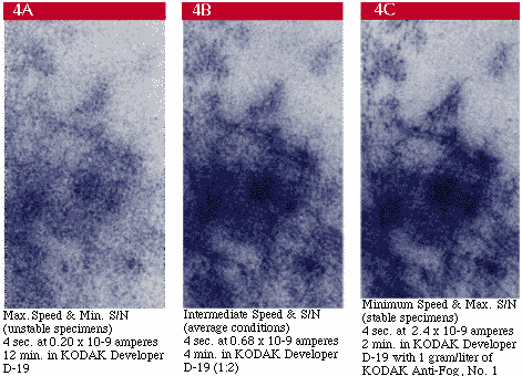
Figure 4: Speed and signal-to-noise characteristics of Electron Image Film may be adjusted to accommodate specimen stability conditions by selecting compensatory exposure and development conditions. These conditions serve as starting points.
In Figure 4, beam current is lowest (fewest electrons) in 4A, intermediate in 4B, and highest (most electrons) in 4C. In the same order, development time and developer activity have been reduced to compensate for the increasing number of electrons collected. In effect, the density contribution per electron that is needed to produce a given density is modified in processing to accommodate the number of absorbed electrons. The goal here, again, is to use more electrons. Collect as many electrons as specimen stability will allow and adjust development conditions to achieve the desired density and contrast.
Causes of Electron Micrography Noise
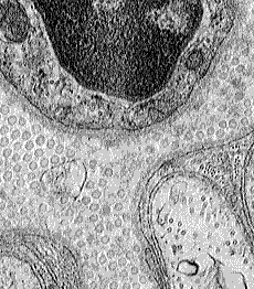
The processes used in electron micrography and ordinary photography are similar in many respects. Both involve exposing a photographic material, processing this material to a negative image, and printing the negative to an enlarged positive print. The primary difference is the exposing radiation-electrons for electron micrography and light for conventional photography. This difference is a critical factor when working with TEMs and photographic techniques, because electrons interact with photographic emulsions in a substantially different manner than do photons. Random fluctuations of electrons in the beam are normal. These fluctuations result in a characteristic grainy appearance in the processed photographic negative. This granular structure (noise) is most readily observed in areas of uniform electron exposure (Figure 5A), and is not due to the inherent photographic grain of the emulsion (Figure 5B) or, necessarily, an indication of instrument instability.


In addition, each incident electron is capable of interacting with a number of silver halide grains along its irregular path through the emulsion, causing them to be developable. Electrons, therefore, efficiently contribute to image density. Combined with the characteristic fluctuations in the beam, however, this efficiency contributes to the granular structure which we recognize as noise in the image.
When light is the exposing radiation, on the other hand, a number of photons must interact with each silver halide grain in order to render it developable. This is primarily due to the energy level of photons (2 to 3 electron volts for visible radiation), which is many times lower than that of electrons in a typical TEM (50,000 to 100,000 electron volts). With photon exposure, the transmittance of a specimen is sampled at a rate that is an order of magnitude greater than with electron exposure. As a result, granularity with photon exposure is reduced to the level of the emulsion itself.
Choose a Region
- Africa
- Asia Pacific
- GCC-TEST
- Europe
- Middle East
- North America
- South America
Africa
Asia Pacific
GCC-TEST
Europe
|
Serbia
Slovakia Slovenia Spain Sweden Switzerland Ukraine United Kingdom |
Middle East
North America
South America
|
São Vicente and Granadina
Suriname Trinidad and Tobago Turk and Caicos Islands Uruguay U.S. Virgin Islands Venezuela Virgin Islands (British) |

