Radiology in the First World War
Reading Time: 4 minutes read
Radiology a hundred years ago was still a heroic endeavor not just for the patients but also for the personnel. Concepts such as protection against the danger of ionizing radiation were largely unknown or ignored.
The horrors of war stimulated a remarkable period of technological development in the then embryonic field of medical imaging.
At the eleventh hour of the eleventh day of the eleventh month in 1918 the armistice was declared to bring to the end one of the most murderous of all conflicts, with an estimated grand total of some 37 million soldiers reported as killed, wounded or missing. The armistice ceremonies, still celebrated in most countries with appropriate solemnity on the 11th of November are always very poignant, but this year, exactly one hundred years after the onset of the First World War they are particularly so.
In the face of the overwhelmingly bleak immensity of the casualty statistics, it is very difficult to unearth any positive aspects but, as so often happens, the sheer force and immediacy of military imperatives actually resulted in positive developments which far outlasted the war itself. Such a situation occurred in the First World War where the overwhelming need for better and more immediate care of the huge numbers of wounded soldiers brought about a step-change in radiology. There was of course a ruthless logic to the need for improved treatment of military wounded in the First World War — the quicker a soldier could be treated, the higher the chance that he would survive and be able to be sent back to the front again.
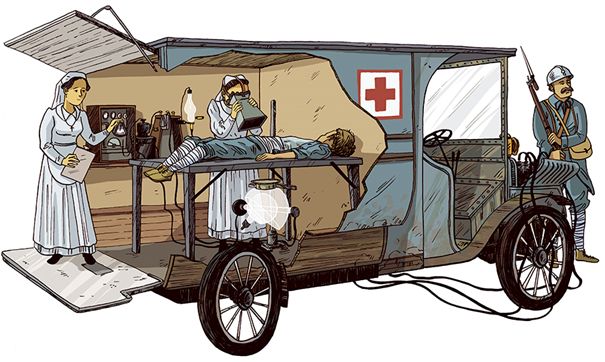
It was difficult and time-consuming to evacuate the thousands of wounded soldiers from the trenches to traditional military hospitals located far behind the lines. The solution was to develop mobile radiology units which could get closer to the battlefields. The French in particular were active in this aspect, which required considerable ingenuity to overcome problems such as those caused by the then fragility of the radiology equipment. Image visualization techniques have changed in the hundred years since the First World War.
Firstly, let’s put things in context. As every modern radiologist knows the birth of radiology occurred in 1895 — so not even twenty years before the First World War — with the discovery in 1895 by WC Röntgen of X-rays and their then mysterious power of being able to visualize the interior of living tissue and organs. By the standard of the time, the news of the invention spread very rapidly thanks to the use of the latest newfangled invention the telegraph (of course at a snail’s pace as compared to modern communication methods of phone internet, Facebook and Twitter, etc.). Interestingly, the military was one of the first to realize the potential of the new X-ray technique. Thus only one year after Röntgen’s ground-breaking discovery, there were already reports of the use of radiology by military surgeons in the Kaiser Wilhelm Academy in Berlin.
Thus, by the time of the onset of the First World War, the potential of radiology for the examination of soldier’s wounds was well recognized by the military authorities. The principal application was brutally simple. With a wounded soldier of a hundred years ago, no one would bother looking for suspicious probably cancerous soft tissue lesions. Instead the primary objective of radiology was to identify the location of metallic foreign bodies such as shrapnel, projectiles etc. lodged within the soldier’s body so that the surgeon could extricate them as quickly as possible. Such metallic foreign bodies are a potential source of infection. The early radiology systems were actually quite good for this purpose — apart from the obvious disadvantage that a radiograph is a two-dimensional projection of a three dimensional volume. Recognizing this, several ingenious solutions were proposed, including the wartime invention by the Frenchman A. Bocage of a rudimentary tomographic system, using the principle of two images of the same patient but taken from different angles, with of course no computer to reassemble the images. The CT scanner as we know it nowadays was still many years off.
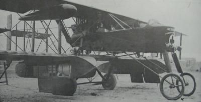
The sheer number of casualties was the stimulus for remarkable technological development. By the end of the war, there even existed air-borne mobile radiology units. Here a converted bomber carries in its under-wing pods a complete radiology station which could be rapidly set up in an inflatable tent also carried in the plane.
In fact as the sheer numbers of wounded soldiers increased dramatically in the early war years, the problems were not so much the basic technique itself, but rather the organization and structure of the military medical services responsible for radiology. The standard model was to have fully equipped military hospitals located far behind the lines. However the difficulty and time taken to evacuate so many wounded soldiers from the front lines to the hospitals meant that by the time they arrived their situation had deteriorated so seriously that medically there was almost nothing to be done.
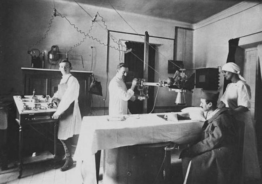
Hence a huge effort was expended into making mobile radiology systems to be able to positioned wherever were the biggest needs. There were however huge problems to be overcome in the development of early mobile radiology units, principally due to the fragility of the early radiology equipment and the need for a supply of electrical power. The abandonment of the traditional horse-drawn cart in favour of the then new technology of automobiles had the advantage of being able to rig up special dynamos run off the motor engine. The French in particular were very active during the first world war in the development of motor driven mobile radiology units. Even Marie Curie and her daughter Irène were actively involved.
The speed at which solutions were introduced to previous problems was quite remarkable and indicative of the urgency of the situation. For example, one problem of mobile radiology units was that under the conditions of war the glass photographic plates used at the time frequently broke. This explains how avidly the invention by Eastman of cellulose-based films coated with photographic emulsions was seized by military radiologists. Likewise the invention of the hot cathode X-ray tube finally solved the short life time of the cold tube. By the end of the war the inventive process reached its peak with the introduction of a Franco-American project of an air-borne radiology unit, where a converted Voisin bomber carried all the equipment necessary to set up a radiology surgical room quickly installed under an inflatable tent.
Despite these advances, one mustn’t become misty – eyed or nostalgic about radiology of the time. Compared to today’s equipment and performance, radiology during the First World War was still a heroic and relatively primitive undertaking. In particular the concept of protection against the dangerous side effects of radiation was effectively non-existent.
Of course the remarkable developments occurring in radiology during the First World War go nowhere near to justifying the carnage which was the underlying impetus for the developments. At best it is a meagre consolation that, in the midst of the colossal human tragedy, a little spark of benefit was found in an embryonic technology that has since grown into today’s indispensable profession.
Alan Barclay, PhD., is the editor of Diagnostic Imaging Europe.